


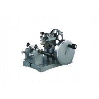


In biomedical laboratories, there is a class of equipment that appears low-key but is almost standard in pathology departments, neuroscience labs, and drug development centers – the cryostat microtome. Unlike ordinary cryostats used for physical material research, the cryostat microtome is specifically designed to cut biological tissues into micron-thick sections at low temperatures for microscopic observation and analysis. In recent years, driven by precision medicine and translational research, the application fields of the cryostat microtome are expanding at an unprecedented pace.
What is a Cryostat Microtome?
Simply put, it is an instrument that combines a freezing chamber with a precision sectioning device. Tissue samples are quickly frozen and hardened inside a constant-temperature chamber at –20°C to –40°C, then cut into sections as thin as 1–100 microns using a high-precision blade, eliminating the need for tedious steps such as paraffin embedding and dehydration. This "freeze-and-cut-on-the-spot" characteristic makes it indispensable in situations requiring rapid diagnosis or preservation of the tissue's original state.
Core Application 1: Rapid Clinical Pathological Diagnosis
In the operating room, time is life. When a cancer patient undergoes tumor removal surgery, the surgeon urgently needs to know whether cancer cells remain at the resection margin – which determines whether a wider excision is necessary. Intraoperative frozen section examination is the "gold standard" tool for this purpose.
The intraoperative frozen section process is: tissue removed during surgery is immediately sent to the pathology department, where a technician freezes, sections, and stains it – the entire process takes only 15–30 minutes. The pathologist then examines the slide under a microscope to determine benign vs. malignant status, margin status, and whether lymph nodes have metastases. According to statistics from the Pathology Department of Peking Union Medical College Hospital, more than 5,000 intraoperative frozen section examinations are completed each year relying on cryostat microtomes, with an accuracy rate exceeding 95%. For surgeries on the thyroid, breast, ovaries, and other sites, this technology greatly reduces the probability of a second operation.
Core Application 2: Neuroscience Research
Brain and spinal cord tissues are soft and rich in lipids; conventional paraffin sections tend to crack or shrink. The cryostat microtome perfectly solves this problem. Researchers can freeze fresh or fixed brain tissue and cut it into thick sections of 20–100 microns for neuroanatomical tracing, immunohistochemistry, or in situ hybridization.
For example, in Alzheimer's disease research, scientists use a cryostat microtome to cut mouse brains into serial sections and then stain with amyloid-beta antibodies to clearly visualize the distribution of senile plaques. Similarly, in stroke models, by combining frozen sections with TTC staining, the volume of cerebral infarction can be accurately measured. It can be said that without the cryostat microtome, the efficiency of modern neuroscience research would be greatly diminished.
Core Application 3: Drug Development and Toxicological Evaluation
Before a new drug can be marketed, its safety and efficacy must be assessed in animals. Pathological examination is a core component of toxicological evaluation. Tissues such as heart, liver, kidney, and lung removed from rats, dogs, or monkeys – some of them need to be sectioned on a cryostat for Oil Red O staining, a special stain that detects fat vacuoles in tissues and is a key indicator of whether a drug causes fatty degeneration. Paraffin sectioning dissolves lipids, so it must be done with a cryostat microtome.
Furthermore, in pharmacodynamic studies of anti-tumor drugs, researchers often rapidly freeze and section xenograft tumors and use Ki-67 or TUNEL staining to assess the drug's effects on tumor proliferation and apoptosis. Frozen sections maximally retain antigen activity, making immunofluorescence signals stronger and more reliable.
Core Application 4: Metabolic Disease and Adipose Tissue Research
Obesity and diabetes are global health challenges. The study of adipose tissue is inseparable from the cryostat microtome. Because lipid droplets inside adipocytes are dissolved by organic solvents during conventional paraffin embedding, leaving empty spaces. Frozen sections combined with Oil Red O or Sudan Black staining can truthfully reveal the size and distribution of lipid droplets.
For example, when studying the transformation between white and brown adipose tissue, researchers freeze-section mouse epididymal fat pads or interscapular brown fat to observe multilocular lipid droplets and UCP1 protein expression. This type of work provides direct histological evidence for the development of weight-loss drugs.
Core Application 5: Botany and Agricultural Science
Plant tissues contain large amounts of water and cell walls, and often have lignified or suberized structures. Conventional paraffin sectioning is time-consuming and difficult. A cryostat microtome can cut fresh plant stems, leaves, or root sections within minutes for observing vascular bundle structure, stomatal distribution, or pathogen infection.
For example, in plant disease resistance research, scientists freeze-section wheat leaves infected with fungi and then stain with fluorescent dyes to directly observe the expansion pathway of hyphae between mesophyll cells under a confocal microscope. This method eliminates dehydration, embedding, and other steps, avoiding morphological changes in the fungus caused by chemical treatment.
Technological Frontiers: The Future Evolution of Cryostat Microtomes
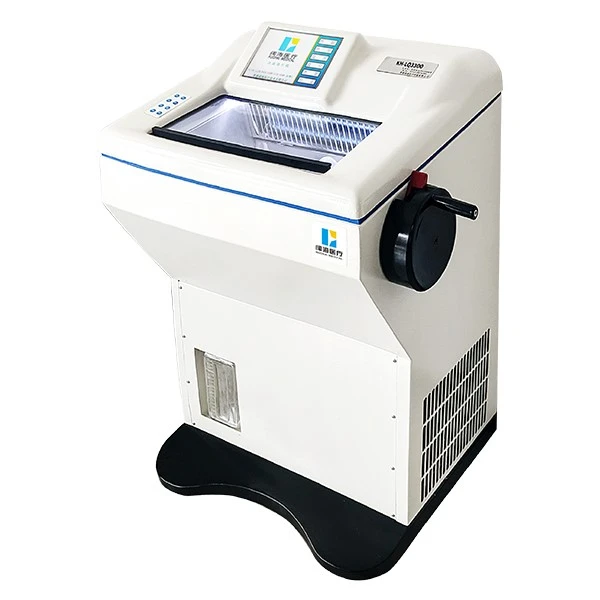
Modern cryostat microtomes are no longer the "simple cold boxes" of the past. New models are equipped with:
Touchscreen intelligent control: can store sectioning parameters (thickness, temperature, anti-roll plate position) for hundreds of tissue types, recallable with one touch.
Motorized sectioning system: ensures uniform section thickness and reduces human error.
UV disinfection and antimicrobial surfaces: prevent operator infection with pathogens, especially valuable for biosafety protection in research involving HIV, tuberculosis, or novel coronaviruses.
Rapid freezing stage (Peltier cooling): can cool tissue to –40°C within 2 minutes, greatly reducing waiting time.
Industry insiders predict that with the rise of spatial transcriptomics, the cryostat microtome will play an even more critical role – because such technologies require capturing mRNA directly on fresh frozen tissue sections, demanding extremely high tissue morphology and nucleic acid integrity. Any fixation or embedding process could cause signal loss.
Conclusion: A "Low-Temperature Knife" Unlocks Countless Mysteries
From life-and-death decisions on the operating table to detailed mapping in brain science; from Oil Red O staining of fat cells to the microscopic world of plant roots and stems – the cryostat microtome has long transcended being a mere instrument, becoming a bridge connecting tissue morphology and molecular information. It allows scientists to peer into the delicate construction of life in a state closest to physiology.
If your laboratory or research institution is considering purchasing or upgrading frozen sectioning equipment, it is worthwhile to look at newer models featuring intelligent temperature control, optimized anti-roll plates, and motorized advance functions. A good "low-temperature scalpel" may well be the starting point for your next major discovery.